Home
/ Plant Cell Diagram Under Light Microscope : Eukaryotic Plant Cell Microscope - Micropedia / Answer the following questions in your exercise book.
Plant Cell Diagram Under Light Microscope : Eukaryotic Plant Cell Microscope - Micropedia / Answer the following questions in your exercise book.
Plant Cell Diagram Under Light Microscope : Eukaryotic Plant Cell Microscope - Micropedia / Answer the following questions in your exercise book.. But at the same time it is interpretive. 1.5 (p.4) which is a diagram of a generalized plant cell. Here's a diagram of a plant cell: Cells consist of cytoplasm enclosed within a membrane, which contains many biomolecules such as proteins and nucleic acids.2 most plant and animal cells are only visible under a light microscope, with dimensions between 1 and 100 micrometres.3 electron microscopy gives a much higher. Cell is a tiny structure and functional unit of a living organism containing various parts known as organelles.
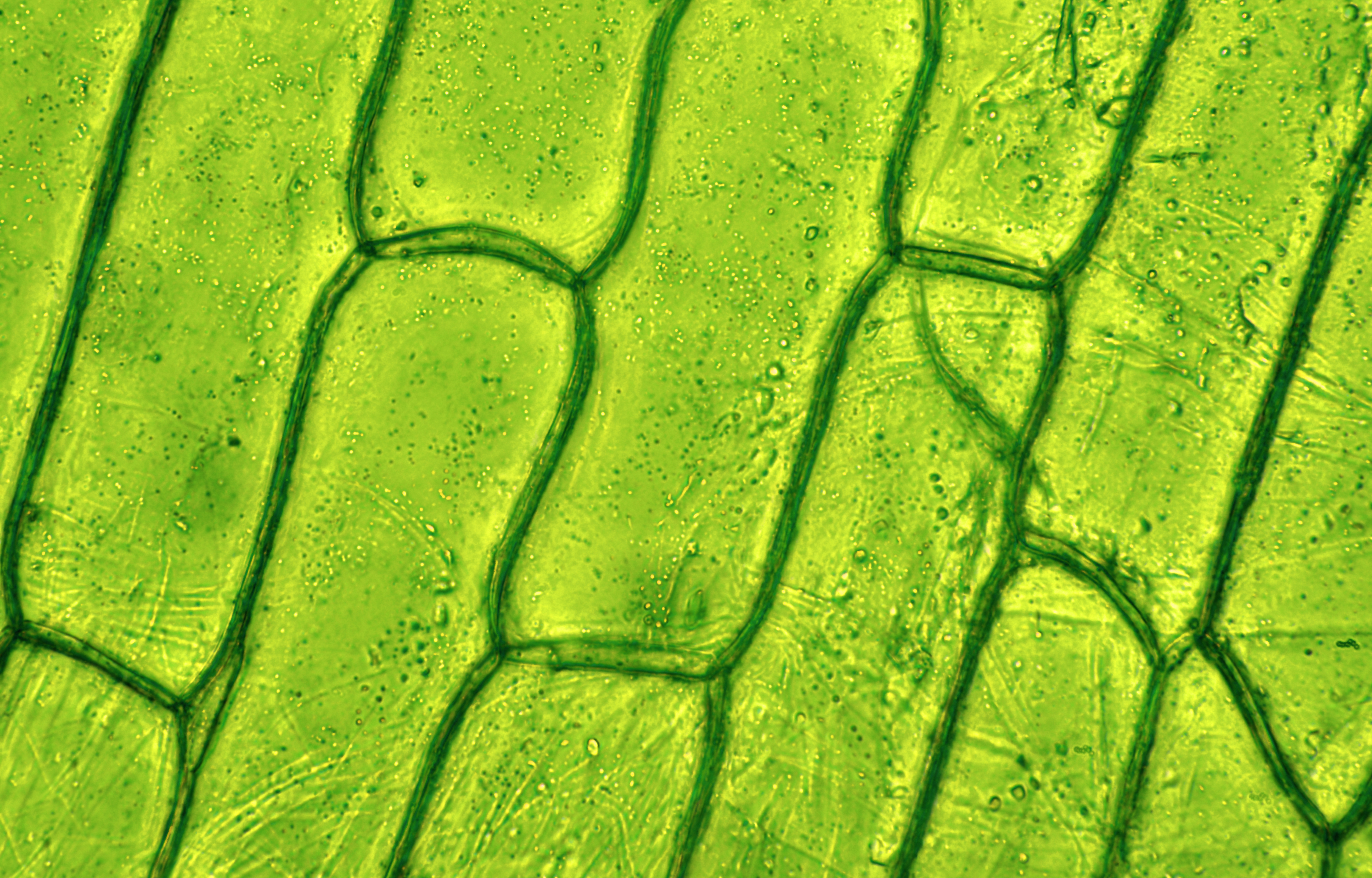
Cells consist of cytoplasm enclosed within a membrane, which contains many biomolecules such as proteins and nucleic acids.2 most plant and animal cells are only visible under a light microscope, with dimensions between 1 and 100 micrometres.3 electron microscopy gives a much higher. Magnification, however, is not the most important issue in microscopy. Robert hooke in 1665 first discovered plant cell. Turn the coarse focus so that the stage is as close to the objective lens as possible. Plant cell surface of leaf under light microscope.
Stomata Under Microscope Labeled - Micropedia from ssl.bigstockimages.com The high resolving power makes the electron microscope a very important research tool in microbiology. In contrast to normal cells, cancer cells often. Learn the structure of animal cell and plant cell under light microscope. Plant cell surface of leaf under light microscope. Study the two diagrams of plant and animal cells below. Plant cells have cell walls, one large vacuole per cell, and chloroplasts, while animal cells will have a cell membrane only. General plant cell as seen under the light microscope. Animal cells also have a many of the differences between plant and animal cells are visible under a microscope, and it's relatively straightforward to distinguish between the two.
Answer the following questions in your exercise book.
Cell is a tiny structure and functional unit of a living organism containing various parts known as organelles. General plant cell as seen under the light microscope. Learn the structure of animal cell and plant cell under light microscope. Purple colored, large epidermal cells of an onion oyster plant cells. A few cell organelles can be seen when a plant cell is viewed under a light microscope. The inner layer is continuous and forms flattened membrane sacs called thylakoids. Plant cell is an eukaryotic cell primarily involved in photosynthesis and having its genomic content some of these differences can be clearly understood when the cells are examined under an electron microscope. Plant cells are the basic unit and building blocks of life in organisms of the kingdom plantae. A micrograph is a photo or digital image taken through a microscope to show a magnified image of a specimen. It also has a very high resolving power. Cells of plant or animal tissue. Cell is a tiny structure and functional unit of a living organism containing various parts known as organelles. 2 units of measurement in cell studies.
In contrast to normal cells, cancer cells often. Resolving power is the ability to distinguish between separate things which are close to each other. We say cells are microscopic because they can only be seen under a microscope. A scale bar has been marked on the drawing, allowing the size of a cell to be estimated. The diagram below is a plant cell as may be seen using a light microscope.
Transverse (A-D, F) and longitudinal (E) light microscope ... from www.researchgate.net In contrast to normal cells, cancer cells often. 1 lab plant and animal cells, light microscopic analysis of leaf cross sections upper, structure of animal cell and plant cell under microscope, vacuole stock photos vacuole stock images alamy, cell structure teaching resources the science teacher. Transport proteins modified by the golgi body outside of the cell. Under ordinary light microscope only few cell organelles like mitochondria, golgi complex however, under electron microscope, several other cytoplasmic organelles such as endoplasmic plastids are present only in plant cells (not in animal cells). Here's a diagram of a plant cell: Plant cells have cell walls, one large vacuole per cell, and chloroplasts, while animal cells will have a cell membrane only. They can be observed easily in a phase contrast microscope under dark field. Image:plant cell seen under electron microscope.
Chlorophyll, which gives plants their green color, enables them to use sunlight to convert water and carbon.
The condenser focuses the light through the object. A few cell organelles can be seen when a plant cell is viewed under a light microscope. Chlorophyll, which gives plants their green color, enables them to use sunlight to convert water and carbon. But at the same time it is interpretive. In truth, there are still features of plant and animal appearance —under a microscope, normal cells and cancer cells may look quite different. Most light microscopes will enlarge a specimen up to 1000 times (1000x) but the electron microscope enlarge the specimen 250. Endoplasmic reticulum studded with ribosomes looks rough under the microscope; A scale bar has been marked on the drawing, allowing the size of a cell to be estimated. The diagram is very clear, and labeled; Microscopy and the interpretation of cell structures. The term 'cell' was coined to describe the small walled units that were observed in the sections they are just visible as small rods or spheres under light microscope. Here's a photo of a plant cell under an electron microscope. Describe and compare the structure of a plant cell with an animal cell, as seen under a light microscope, limited to cell wall, nucleus, cytoplasm, chloroplasts, vacuoles and location of the cell membrane.
Purple colored, large epidermal cells of an onion oyster plant cells. Cheek cell(animal cell) onion cell(plant cell). It also has a very high resolving power. Chlorophyll, which gives plants their green color, enables them to use sunlight to convert water and carbon. To use a light microscope to examine animal or plant cells.
How plant cells neutralize the potential for self-harm ... from carnegiescience.edu Here's a photo of a plant cell under an electron microscope. Cell organelles are generally colourless and must be stained to see them e.g. Plant cell is an eukaryotic cell primarily involved in photosynthesis and having its genomic content some of these differences can be clearly understood when the cells are examined under an electron microscope. In contrast to normal cells, cancer cells often. Here's a diagram of a plant cell: Transport proteins modified by the golgi body outside of the cell. General animal cell as seen under the light microscope. The diagram below is a plant cell as may be seen using a light microscope.
This shows a generalized animal cell under a light microscope.
1 lab plant and animal cells, light microscopic analysis of leaf cross sections upper, structure of animal cell and plant cell under microscope, vacuole stock photos vacuole stock images alamy, cell structure teaching resources the science teacher. (ii) name another type of microscope that gives. Cells of plant or animal tissue. Study the two diagrams of plant and animal cells below. This shows a generalized animal cell under a light microscope. Most light microscopes will enlarge a specimen up to 1000 times (1000x) but the electron microscope enlarge the specimen 250. Microscopy and the interpretation of cell structures. Learn the structure of animal cell and plant cell under light microscope. See how a generalized structure of an animal cell and plant cell look with labeled diagrams. We say cells are microscopic because they can only be seen under a microscope. The diagram is very clear, and labeled; Purple colored, large epidermal cells of an onion oyster plant cells. Cell is a tiny structure and functional unit of a living organism containing various parts known as organelles.
Share :
Post a Comment
for "Plant Cell Diagram Under Light Microscope : Eukaryotic Plant Cell Microscope - Micropedia / Answer the following questions in your exercise book."


Post a Comment for "Plant Cell Diagram Under Light Microscope : Eukaryotic Plant Cell Microscope - Micropedia / Answer the following questions in your exercise book."