Home
/ Electron Microscope Image Of Animal Cell : Microscope Cell Images Animal Cells All Living Things Are Made Up Of One Or More Cells Cells Are The Basic Units Of Structure And Function In Organisms Ppt Download / See how sem cell images although the very first electron microscopy (em) images of eukaryotic cells were attributed in 1945, it was the ruska family that not only developed.
Electron Microscope Image Of Animal Cell : Microscope Cell Images Animal Cells All Living Things Are Made Up Of One Or More Cells Cells Are The Basic Units Of Structure And Function In Organisms Ppt Download / See how sem cell images although the very first electron microscopy (em) images of eukaryotic cells were attributed in 1945, it was the ruska family that not only developed.
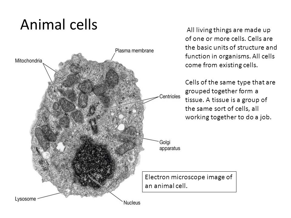
Electron Microscope Image Of Animal Cell : Microscope Cell Images Animal Cells All Living Things Are Made Up Of One Or More Cells Cells Are The Basic Units Of Structure And Function In Organisms Ppt Download / See how sem cell images although the very first electron microscopy (em) images of eukaryotic cells were attributed in 1945, it was the ruska family that not only developed.. Cell structure i nucleus medical media. This is the ability to see two points as two points, rather. A cell is a very tiny structure which exists in living bodies. The shorter the wavelength of the illumination, the better the. Collagen fibrils were found investing the perineurial cells, and occasional.
However, light microscopes form real colour images and can be used to watch living processes occur in microscopic detail, while electron microscopes cannot be used to study living cells. An electron microscope is a microscope that uses a beam of accelerated electrons as a source of illumination. Electron microscopes use electron beams focused by electromagnets to magnify and resolve microscopic specimens. Both plant and animal cells are surrounded by a cell membrane composed of lipids and proteins. The colours are added afterwards using specialised computer software.
A Typical Animal Cell As Seen In An Electron Microscope Medical Ima from image.slidesharecdn.com Animals/wildlife buildings/landmarks backgrounds/textures business/finance education food and drink health care holidays objects industrial art nature people religion science technology download this image now with a free trial. Cell structure i nucleus medical media. Electron microscopes use electron beams focused by electromagnets to magnify and resolve microscopic specimens. Cells of plant or animal tissue. They participate in cell reproduction in in addition, and image viewed through a microscope is much more detailed, and it shows all of the. The colours are added afterwards using specialised computer software. A cell is a very tiny structure which exists in living bodies. Confocal microscopy image of a young leaf of thale cress, with one marker outlining the cells and other markers indicating young cells of the stomatal lineage.
See more ideas about electron microscope images, electron microscope, microscopic photography.
See more ideas about electron microscope images, electron microscope, microscopic photography. Membrane bound nucleus is present in both with a transmission electron microscope (tem) and generic contrast staining (osmium, uranyl, lead) of a section through a cell you will not only see the. Visible are neural cell bodies, complete with. Plus, get full access to a library of over 316 million images. They participate in cell reproduction in in addition, and image viewed through a microscope is much more detailed, and it shows all of the. In this microscope, images are produced from the interaction between the prepared samples in the vacuum. Cells of plant or animal tissue. Most cells are very small, and their structures can only be seen by using a microscope. Scanning electron microscope images reveal hidden horror and beauty. Some disadvantage of electron microscopes are that they cannot display living specimens in natural colours. Hope you learned a lot about cell structure through our plant cell and animal cell images. Electron microscopes use electron beams focused by electromagnets to magnify and resolve microscopic specimens. This is an electron microscope image showing part of the rough endoplasmic.
This is an electron microscope image showing part of the rough endoplasmic. Confocal microscopy image of a young leaf of thale cress, with one marker outlining the cells and other markers indicating young cells of the stomatal lineage. When you look at animal or plant cells under the electron microscope, you can see a lot more detail. The animal cell is more. All images are 100% editable in powerpoint.
Electron Microscopes Cell Structure Edexcel Gcse Combined Science Revision Edexcel Bbc Bitesize from ichef.bbci.co.uk Category:electron microscope images (en) categoría de wikimedia (es); This is the ability to see two points as two points, rather. Collagen fibrils were found investing the perineurial cells, and occasional. Learn vocabulary, terms and more with flashcards in the cells of animals and some fungi and algae. 50 amazing things under electron microscope sem images . Electron microscope images scanning electron micrograph microscopic photography macro photography science images microscopic images macro electron microscope reveals strange features of familiar items. Recent experimentation has been aimed at utilizing animal cells. The shorter the wavelength of the illumination, the better the.
Plant, animal and bacterial cells have smaller components each with a the ability to see greater detail in an image depends on the resolution or resolving power.
See how sem cell images although the very first electron microscopy (em) images of eukaryotic cells were attributed in 1945, it was the ruska family that not only developed. Visible are neural cell bodies, complete with. This is the ability to see two points as two points, rather. Level suitable for as biology. Category:electron microscope images (en) categoría de wikimedia (es); The animal cell is more. Electron microscopes use accelerated electron beams (as opposed to visible light in a light microscope) to create images of magnification as here is an electron micrograph of an animal cell with the labels superimposed: However, light microscopes form real colour images and can be used to watch living processes occur in microscopic detail, while electron microscopes cannot be used to study living cells. Scanning electron microscopy is used in cell biology research for imaging cellular organelles and surface topography. The shorter the wavelength of the illumination, the better the. Electron microscope images scanning electron micrograph microscopic photography macro photography science images microscopic images macro electron microscope reveals strange features of familiar items. Start studying cell structure & microscopes. Anatomy_and_physiology_of_animals_animal_cell_electron_microscope.jpg (557 × 540 pixels, file size:
Scanning electron micrographs of all kind of small animals. For images showing electron microscopes see category:electron microscopes. Unlike the eukaryotic cells of plants and fungi, animal cells do not have a cell wall. The animal cell is more. Animals/wildlife buildings/landmarks backgrounds/textures business/finance education food and drink health care holidays objects industrial art nature people religion science technology download this image now with a free trial.
Microscope Cell Images Animal Cells All Living Things Are Made Up Of One Or More Cells Cells Are The Basic Units Of Structure And Function In Organisms Ppt Download from images.slideplayer.com See more ideas about electron microscope images, electron microscope, microscopic photography. 50 amazing things under electron microscope sem images . Learn vocabulary, terms and more with flashcards in the cells of animals and some fungi and algae. When you look at animal or plant cells under the electron microscope, you can see a lot more detail. Electron microscopes use accelerated electron beams (as opposed to visible light in a light microscope) to create images of magnification as here is an electron micrograph of an animal cell with the labels superimposed: All images are 100% editable in powerpoint. You see that many features are in common. However, light microscopes form real colour images and can be used to watch living processes occur in microscopic detail, while electron microscopes cannot be used to study living cells.
Start studying cell structure & microscopes.
Electron microscopes use electron beams focused by electromagnets to magnify and resolve microscopic specimens. Level suitable for as biology. Most compound microscopes today have an illuminator built into the base. Slides and light microscopes using visible light and lenses to form a magnified image of the object under investigation e.g. This is the ability to see two points as two points, rather. Membrane bound nucleus is present in both with a transmission electron microscope (tem) and generic contrast staining (osmium, uranyl, lead) of a section through a cell you will not only see the. This is an electron microscope image showing part of the rough endoplasmic. Scanning electron microscope images reveal hidden horror and beauty. As the wavelength of an electron can be up to 100. Introduction to microscopes and how they work. For images showing electron microscopes see category:electron microscopes. When cells are imaged in the microscope, even for short periods of time, these same medium conditions must be carefully reproduced in the the pinnacle of live cell imaging chambers effectively combines a cell culture incubator with an inverted microscope to provide almost total control of the. Scanning electron microscopy is used in cell biology research for imaging cellular organelles and surface topography.
Light and electron microscopes allow us to see inside cells animal cell electron microscope. Start studying cell structure & microscopes.
Share :
Post a Comment
for "Electron Microscope Image Of Animal Cell : Microscope Cell Images Animal Cells All Living Things Are Made Up Of One Or More Cells Cells Are The Basic Units Of Structure And Function In Organisms Ppt Download / See how sem cell images although the very first electron microscopy (em) images of eukaryotic cells were attributed in 1945, it was the ruska family that not only developed."


Post a Comment for "Electron Microscope Image Of Animal Cell : Microscope Cell Images Animal Cells All Living Things Are Made Up Of One Or More Cells Cells Are The Basic Units Of Structure And Function In Organisms Ppt Download / See how sem cell images although the very first electron microscopy (em) images of eukaryotic cells were attributed in 1945, it was the ruska family that not only developed."