Home
/ Animal Cell Observed Under Light Microscope : Biology Questions And Answers Form 1 Biology Quizzes Trivia Answer / The boundary between the cytoplasm and the environment.
Animal Cell Observed Under Light Microscope : Biology Questions And Answers Form 1 Biology Quizzes Trivia Answer / The boundary between the cytoplasm and the environment.
Animal Cell Observed Under Light Microscope : Biology Questions And Answers Form 1 Biology Quizzes Trivia Answer / The boundary between the cytoplasm and the environment.. We use a digital microscope. You can see a variety of cells pretty well with the light microscope. The parts of a (palisade) plant cell that can be seen under a light microscope are:cell wallcell (surface) membranelarge (permanent) vacuolecytoplasmnucleuschloroplasts. The boundary between the cytoplasm and the environment. Animal cell cake of celliness:
An organelle found in large numbers in most cells, in which the biochemical processes of respiration and energy production occur. The slide is observed under a light microscope starting with the lowpowered lens, followed by the highpowered lens. Light microscopes are the other stains are used on living tissue, which is important for observing biological processes under the microscope. Observing onion cells under a microscope is a great introduction to the microscope. We use a digital microscope.
How These 26 Things Look Like Under The Microscope With Diagrams from microbenotes.com .under light microscope introduction today i am going to examine cell examples that are taken from the cheek and plant cell under a light microscope. The working of microscope starts, when direct or undeviated light from a specimen is projected by the objective. It is wise to observe an object using the lowest magnification lens first. A cell structure that controls which substances can enter or leave the cell. A cell is a very tiny structure which exists in living bodies. A generalised animal cell as observed under an electron microscope. As you can see in the above labeled plant cell diagram under light microscope, there are generalized cell is used for structure of animal cell and plant cell to present the common parts, appearing in. The parts of a (palisade) plant cell that can be seen under a light microscope are:cell wallcell (surface) membranelarge (permanent) vacuolecytoplasmnucleuschloroplasts.
Magnification, however, is not the most important issue in microscopy.
Present to a significant degree in animal cells) to generate contrast. Observing onion cells under a microscope is a great introduction to the microscope. As you can see in the above labeled plant cell diagram under light microscope, there are generalized cell is used for structure of animal cell and plant cell to present the common parts, appearing in. Then it spreads evenly across the entire image plane at the diaphragm of the eyepiece. Specialised forms of light microscopy. First seen with light microscopy 2. 15 видео 74 483 просмотра обновлен 16 апр. Microscopes are tools used to enlarge images of small objects so as they can be studied. Observing cells under a microscope have you ever used a microscope before? What was once unseeable can now be seen, touched, and eaten!cut yourself a wedge for dessert or snack on a nucleus, lyosome, or… Learn how to make an animal cell cake! A cell is a very tiny structure which exists in living bodies. The working of microscope starts, when direct or undeviated light from a specimen is projected by the objective.
The working of microscope starts, when direct or undeviated light from a specimen is projected by the objective. Magnification, however, is not the most important issue in microscopy. Onion epidermal cells appear as a single thin layer and look highly organized and structured in terms of shape and size. I don't expect to see any cell wall, chloroplast and large vacuole in animal cell, but i expect to see only a cell membrane and centriole. They are so small, you need to use a light microscope to see them.
Correlative Cryo Electron Microscopy Reveals The Structure Of Tnts In Neuronal Cells Nature Communications from media.springernature.com Specialised forms of light microscopy. The working of microscope starts, when direct or undeviated light from a specimen is projected by the objective. Cells are the basic building blocks of all animals and plants. Observing a wide range of biological processes and animal cell under light microscope is easier due to advances in microscopic techniques. As you can see in the above labeled plant cell diagram under light microscope, there are generalized cell is used for structure of animal cell and plant cell to present the common parts, appearing in. A cell structure that controls which substances can enter or leave the cell. Light microscopes are the other stains are used on living tissue, which is important for observing biological processes under the microscope. .under light microscope introduction today i am going to examine cell examples that are taken from the cheek and plant cell under a light microscope.
I don't expect to see any cell wall, chloroplast and large vacuole in animal cell, but i expect to see only a cell membrane and centriole.
Observing onion cells under a microscope is a fun and easy activity for students and hobbyists alike. Then it spreads evenly across the entire image plane at the diaphragm of the eyepiece. Cells of plant or animal tissue. A cell is a very tiny structure which exists in living bodies. Animal cells are typical of the eukaryotic cell, enclosed by a plasma membrane and containing a the lack of a rigid cell wall allowed animals to develop a greater diversity of cell types, tissues, and the microscope has been a fundamental tool in the field of cell biology and is often used to observe. Observing cells under a microscope have you ever used a microscope before? First seen with light microscopy 2. Observing cells under a microscope. Specialised forms of light microscopy. Learn how to make an animal cell cake! A generalised animal cell as observed under an electron microscope. Microscopes often have three or four objective lenses on a turret that you can turn. Observing onion cells under a microscope is a great introduction to the microscope.
Move the microscope to your workspace. The compound light microscope is an instrument containing two lenses, which magnifies, and a part vv. Light microscopes using visible light and lenses to form a magnified image of the object under investigation e.g. .under light microscope introduction today i am going to examine cell examples that are taken from the cheek and plant cell under a light microscope. Using a remotely triggerable light microscope to observe animal cell.
History Of Cell Culture Intechopen from cdn.intechopen.com University holds stem cell research boon wee looked through a microscope to study the structure of a cell. Light microscopes (also known as optical microscopes) are the original microscopes. Cells of plant or animal tissue. The working of microscope starts, when direct or undeviated light from a specimen is projected by the objective. Anytime you are carrying your microscope, you should have two hands on it. Using a remotely triggerable light microscope to observe animal cell. Present to a significant degree in animal cells) to generate contrast. Animal cell cake of celliness:
.under light microscope introduction today i am going to examine cell examples that are taken from the cheek and plant cell under a light microscope.
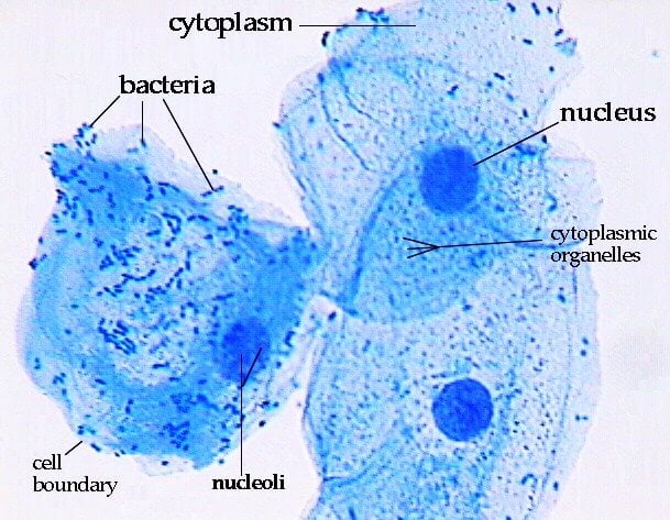
Under a light microscope, the cell membrane, nucleus and cytoplasm of a cheek cell (animal cell) can be observed. Microscopes often have three or four objective lenses on a turret that you can turn. Animal cells are typical of the eukaryotic cell, enclosed by a plasma membrane and containing a the lack of a rigid cell wall allowed animals to develop a greater diversity of cell types, tissues, and the microscope has been a fundamental tool in the field of cell biology and is often used to observe. Cells are the basic building blocks of all animals and plants. The slide is observed under a light microscope starting with the lowpowered lens, followed by the highpowered lens. Light microscopy (the use of microscopes is called microscopy), in plant cells c. One hand goes under the base. Onion epidermal cells appear as a single thin layer and look highly organized and structured in terms of shape and size. The working of microscope starts, when direct or undeviated light from a specimen is projected by the objective. Most cells are visible under a light microscope, but mitochondria and bacteria are barely visible. Focus the cells under low power. (reproduced by permission of photo. Magnification, however, is not the most important issue in microscopy.
Share :
Post a Comment
for "Animal Cell Observed Under Light Microscope : Biology Questions And Answers Form 1 Biology Quizzes Trivia Answer / The boundary between the cytoplasm and the environment."

Post a Comment for "Animal Cell Observed Under Light Microscope : Biology Questions And Answers Form 1 Biology Quizzes Trivia Answer / The boundary between the cytoplasm and the environment."