Animal Cell Diagram Vesicle : Animal Cell Diagram Stock Photo Alamy / To help you do this, i've created a printable animal cell diagram.. Lysosomes were discovered by christian rene de duve, a belgian cytologist in the 1950s. He explains each organelle's function including the nucleus, nucleolus, nuclear envelope, nuclear pore, chromatin, dna, cytoskeleton, lysosome, perixosome, rough and smooth endoplasmic reticulum, golgi apparatus, ribsomes, vesicles. After completing this section, you should know: This is where the digestion of cell nutrients takes place. It is also known as cell vesicles;
The cell membrane, or plasma membrane, is a biological membrane lipids are known to spontaneously form bilayered vesicles in water, and could have preceded rna. Under the microscope, an animal cell shows many different parts called organelles, that work together to keep the cell functional. Macromolecules like proteins and rna pass through. Plant and animal cells have several differences and similarities. I spelt it wrong in the diagram, sorry).
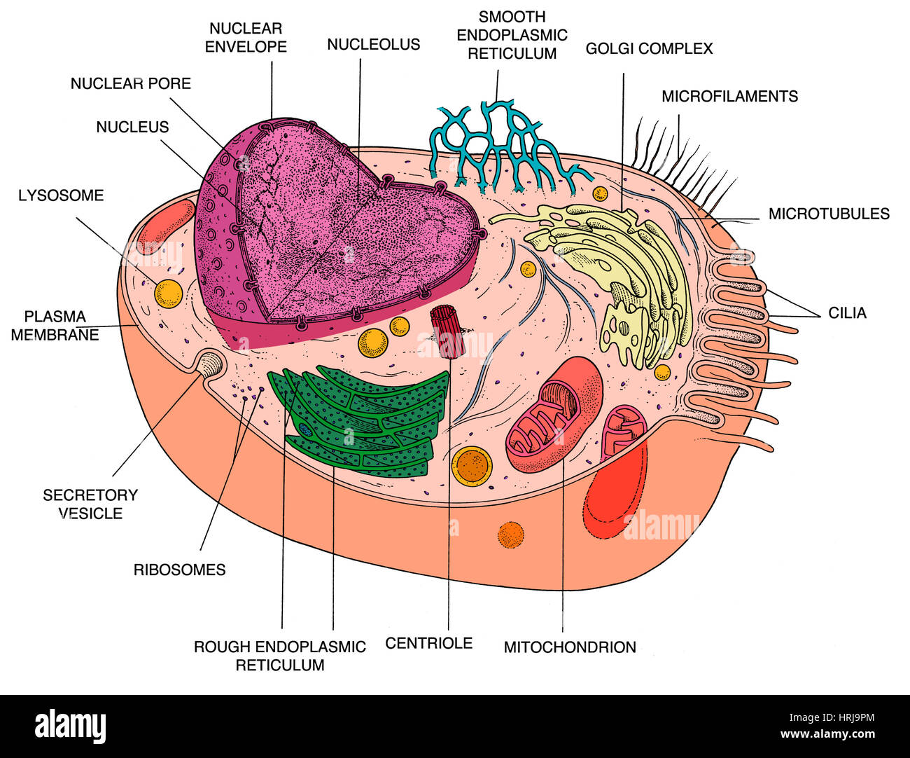
Animal Cell Diagram Stock Photo Alamy from c8.alamy.com After completing this section, you should know: To help you do this, i've created a printable animal cell diagram. Cell organelles structure and parts. Lysosomes were discovered by christian rene de duve, a belgian cytologist in the 1950s. The nuclear pores have small openings that allow the transportation of molecules between the nucleus and cytoplasm. The diagram, like the one above, will include labels of the major parts of an animal cell including the cell membrane, nucleus, ribosomes, mitochondria, vesicles, and cytosol. The role and function of the plasma membrane; The largest organelle within the cell.
The nuclear pores have small openings that allow the transportation of molecules between the nucleus and cytoplasm.
The cell membrane, or plasma membrane, is a biological membrane lipids are known to spontaneously form bilayered vesicles in water, and could have preceded rna. 5th grade science and biology. The number of cells in plants and animals varies from species to species; It is an important cell organelle, which stores and modify proteins for some definite work and transport to vesicles are passive cellular containers — some liquid, enclosed by a membrane — with the inside separated from the cytosol, allowing a wide. Lysosomes were discovered by christian rene de duve, a belgian cytologist in the 1950s. Lets us discuss the animal cell, types of an animal cell, animal cell diagram, its structure. The vacuoles and vesicles should be smaller than the mitochondria. An animal cell ranges in size from 10 to 30 µm. Round organelles surrounded by a membrane and containing digestive enzymes. Cell membrane is made up of lipids and proteins and forms a barrier between the extracellular liquid. Bound ribosome nucleolus rough er (endoplasmic reticulum). Vesicles are compartments formed by a lipid bilayer separating its contents from the cytoplasm or a in each cell they have a distinct function and the same cell can have different types of vesicles intracellular vesicles involved in digestion. A system of flattened membranes called cisternae (mainpoint:
Cell organelles structure and parts. Nucleus smooth er (no ribosomes) centrioles(2). It is also known as cell vesicles; Bound ribosome nucleolus rough er (endoplasmic reticulum). I spelt it wrong in the diagram, sorry).
Animal Cell Definition Structure Parts Functions And Diagram from microbenotes.com It has detailed diagram of lipid bilayer cell membrane. Lets us discuss the animal cell, types of an animal cell, animal cell diagram, its structure. Plant cell and animal cell fall under eukaryotic type. That cells can be of different shapes and sizes. Start studying animal and plant cells. They are used for transport into the cell and will be found outside the cell. Printable animal cell diagram to help you learn the organelles in an animal cell in preparation for your test or quiz. Cell membrane cytoskeleton each animal cell consists of a membrane which is a.
Bound ribosome nucleolus rough er (endoplasmic reticulum).
Learn vocabulary, terms and more with flashcards, games the nucleus, in this diagram, is the yellow part in the middle. It is an important cell organelle, which stores and modify proteins for some definite work and transport to vesicles are passive cellular containers — some liquid, enclosed by a membrane — with the inside separated from the cytosol, allowing a wide. The role and function of the plasma membrane; Animal cell structures, functions & diagrams. Start studying animal and plant cells. Lets us discuss the animal cell, types of an animal cell, animal cell diagram, its structure. Macromolecules like proteins and rna pass through. For example, animal cells do not have a cell wall or chloroplasts but plant cells do. This is where the digestion of cell nutrients takes place. Cell organelles structure and parts. All organisms are made up of cells (or in some cases, a single cell). Animal cells consist of an outer cell membrane filled with cytoplasm and microscopic organelles. An animal cell ranges in size from 10 to 30 µm.
A system of flattened membranes called cisternae (mainpoint: It has detailed diagram of lipid bilayer cell membrane. Learn vocabulary, terms and more with flashcards, games the nucleus, in this diagram, is the yellow part in the middle. It is an important cell organelle, which stores and modify proteins for some definite work and transport to vesicles are passive cellular containers — some liquid, enclosed by a membrane — with the inside separated from the cytosol, allowing a wide. I spelt it wrong in the diagram, sorry).
Retromer And The Trafficking Of Synaptic Vesicle Proteins A Download Scientific Diagram from www.researchgate.net A tour of the animal cell by biology professor dr. Learn vocabulary, terms and more with flashcards, games the nucleus, in this diagram, is the yellow part in the middle. Firstly golgi apparatus (vesicles) is not a cell. The nuclear pores have small openings that allow the transportation of molecules between the nucleus and cytoplasm. All the living organisms are made up of cells and it is the smallest unit • extracellular vesicles: Nucleus smooth er (no ribosomes) centrioles(2). They are used for transport into the cell and will be found outside the cell. Lets us discuss the animal cell, types of an animal cell, animal cell diagram, its structure.
Macromolecules like proteins and rna pass through.
Cell membrane cytoskeleton each animal cell consists of a membrane which is a. It has detailed diagram of lipid bilayer cell membrane. A tour of the animal cell by biology professor dr. Cell organelles structure and parts. For example, animal cells do not have a cell wall or chloroplasts but plant cells do. Diagram showing golgi bodies found in animal cells. The cell membrane, or plasma membrane, is a biological membrane lipids are known to spontaneously form bilayered vesicles in water, and could have preceded rna. The vacuoles and vesicles should be smaller than the mitochondria. Plant and animal cells have several differences and similarities. They are commonly seen in both eukaryotic. Under the microscope, an animal cell shows many different parts called organelles, that work together to keep the cell functional. Animal cells have some organelles not found in the plant cell such as the cytoskeleton, flagalla and cilia. Printable animal cell diagram to help you learn the organelles in an animal cell in preparation for your test or quiz.
Share :
Post a Comment
for "Animal Cell Diagram Vesicle : Animal Cell Diagram Stock Photo Alamy / To help you do this, i've created a printable animal cell diagram."


Post a Comment for "Animal Cell Diagram Vesicle : Animal Cell Diagram Stock Photo Alamy / To help you do this, i've created a printable animal cell diagram."